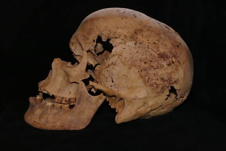
Scientists led by Edgard Camarós, a paleopathologist at the University of Santiago de Compostela in Spain, were studying an approximately 4,600-year-old Egyptian skull when they found signs of brain cancer and its treatment.
There was an uncomfortable silence in the room, because we knew what we had just discovered, Dr. Camarós said.
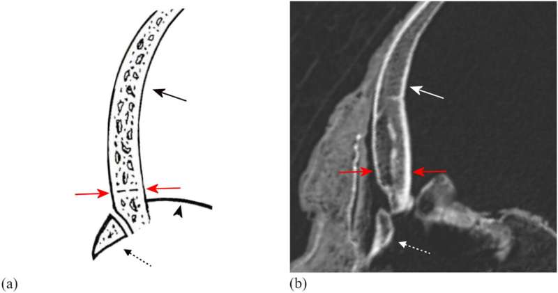
Using a microscope, he and Tatiana Tondini of the University of Tübingen in Germany and Albert Isidro of the University Hospital Sagrat Cor in Spain, the studys other authors, found cut marks around the skulls edges surrounding dozens of lesions that earlier researchers had linked to metastasized brain cancer. The shape of the cuts indicated that they had been made with a metal tool. This discovery, reported in a study published Wednesday in the journal Frontiers in Medicine, suggests that ancient Egyptians studied brain cancer using surgery. If the cuts were made while the person was alive, they may have even attempted to treat it.
The new discovery not only expands scientific knowledge of Egyptian medicine, it may also push back the timeline of humanitys documented attempts to treat cancer by up to 1,000 years.
Cancer has bedeviled humans for as long as we have existed, and it even afflicted life on Earth long before.
Cancer is as old as time, Dr. Camarós said. Even dinosaurs suffered from cancer.
Paleopathologists like Dr. Camarós study the evolution of a disease as well as attempts to understand or treat it. For example, we know that humans in prehistory were afflicted with cancers that no longer exist. He and his colleagues hope that unraveling cancers shifting nature over millenniums may reveal information that can help design treatments for today.
While cancer was probably not well understood, medicine in Egypt was advanced compared with much of the ancient world. An Egyptian document called the Edwin Smith Papyrus, which was written roughly 3,600 years ago, refers to what some researchers believe is a cancer case. That text describes a grave disease for which there was no treatment.
People in ancient Egypt also operated on skulls in other ways. Dr. Camaróss team also reports in the study that they found evidence of successful treatment for a traumatic injury on another cranium, this one 2,600 years old.
Casey L. Kirkpatrick, a bioarchaeologist and a postdoctoral researcher at the Max Planck Institute for Evolutionary Anthropology in Germany, said the new paper presents the first physical evidence of possible cancer treatment by ancient Egyptians.
And by documenting additional ancient historical evidence of the disease, Dr. Kirkpatrick said the study had another benefit.
It can also remind us that cancer is not a modern disease, she said, which might help to relieve some guilt in those currently affected by cancer who are concerned about the role that their lifestyle played in its development.
Just as cancer treatment was a frontier for ancient Egyptians, exploration of the deep past by modern researchers is fraught with uncertainty. The researchers say it is impossible to determine whether the skulls surgical markings were made before death suggesting treatment or after. Many cancers also arise in soft tissues, leaving bones unaffected. This presents a challenge for modern scientists because bones are all that typically survive in the fossil record.
Despite these obstacles, Dr. Camarós said the new discovery gave scientists a fresh perspective on what to look for. He plans to search for similar evidence in ancient sites in Kenya next.
Im sure this is just one example, he said. Posted by BrandonP (Member # 3735) on :
This is an amazing find!
Posted by Djehuti (Member # 6698) on :
^ Because the Egyptians seemed to have had an extensive knowledge of medicine including anatomy and pathology due to their surgical knowledge, it should be no surprise that they made attempts to study and treat such diseases.
I find stories like this fascinating since I myself work in medical pathology.
Oldest Evidence Of Breast Cancer Seen In Ancient Egyptian Skeleton(2015) Archaeologists say they may have found the world's oldest case of breast cancer in a skeleton unearthed recently in Egypt -- a reminder that cancer is not just a modern disease. The skeleton, believed to be that of an adult woman, was unearthed by Spanish researchers working at the Qubbet el-Hawa archaeological site west of Aswan, Egypt. The bones date back 4,200 years and bear signs of "the typical destructive damages provoked by the extension of a breast cancer as a metastasis in the bones," according to a written statement issued by Egypt's Ministry of Antiquities.
^ You can see the bone lesions from the metastatic breast cancer cells.
Who was this unfortunate woman? Evidence suggests she was an aristocrat who lived on the Nile River island of Elephantine during Egypt's 6th Dynasty.
This isn't the first time researchers have found evidence of cancer in ancient times. Last March, archaeologists discovered a 3,000-year-old skeleton with metastatic cancer in a tomb in modern Sudan. And last October, a new MRI analysis of a Siberian mummy showed the "ice princess" likely suffered from breast cancer 2,500 years ago--and used medical marijuana to cope with the disease.
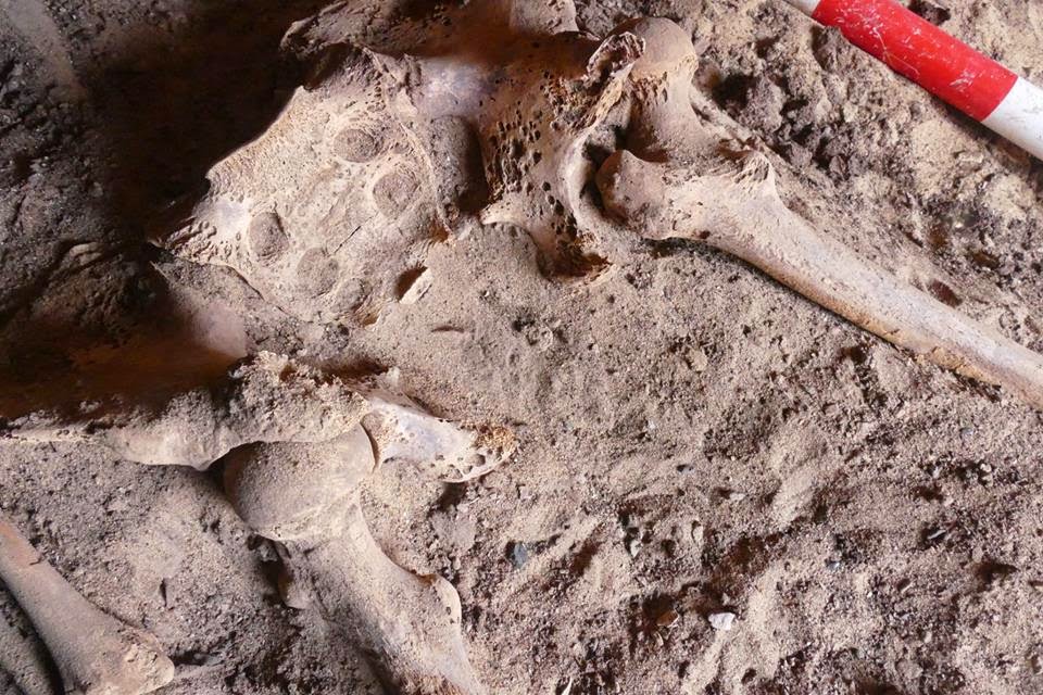
About that finding in Sudan: Ancient Skeleton Found In Sudan Called Earliest Example Of Human With Metastatic Cancer: Ancient Tomb Yields Remains Of Earliest Cancer Patient (2014) British archaeologists have found what they say is the world's oldest complete example of a human being with metastatic cancer and hope it will offer new clues about the now common and often fatal disease.
Researchers from Durham University and the British Museum discovered the evidence of tumours that had developed and spread throughout the body in a 3,000-year-old skeleton found in a tomb in modern Sudan in 2013.
Analysing the skeleton using radiography and a scanning electron microscope, they managed to get clear imaging of lesions on the bones which showed the cancer had spread to cause tumours on the collar bones, shoulder blades, upper arms, vertebrae, ribs, pelvis and thigh bones.
"Insights gained from archaeological human remains like these can really help us to understand the evolution and history of modern diseases," said Michaela Binder, a Durham PhD student who led the research and excavated and examined the skeleton.
"Our analysis showed that the shape of the small lesions on the bones can only have been caused by a soft tissue cancer ... though the exact origin is impossible to determine through the bones alone."
Despite being one of the world's leading causes of death today, cancer is virtually absent in archaeological records compared to other diseases - and that has given rise to the idea that cancers are mainly attributable to modern lifestyles and to people living for longer.
According to the World Health Organisation's cancer research agency, new cancer cases rose to an estimated 14 million a year in 2012, a figure seen rising to 22 million within the 20 years.
Yet these new findings, published in the Public Library of Science journal PLOS ONE on Monday, suggest cancer is not only a modern disease, but was around in the Nile Valley even in ancient times.
Binder said the discovery should help scientists explore the underlying causes of cancer in ancient populations and give fresh clues about the evolution of cancer in the past.
Ancient DNA analysis of skeletons and mummies with evidence of cancer can be used to detect mutations in specific genes that are known to be associated with particular types of cancer.
The skeleton is of an adult male estimated to be between 25- and 35-years-old when he died. It was found at the archaeological site of Amara West in northern Sudan, on the Nile, 750 km downstream from the capital Khartoum.
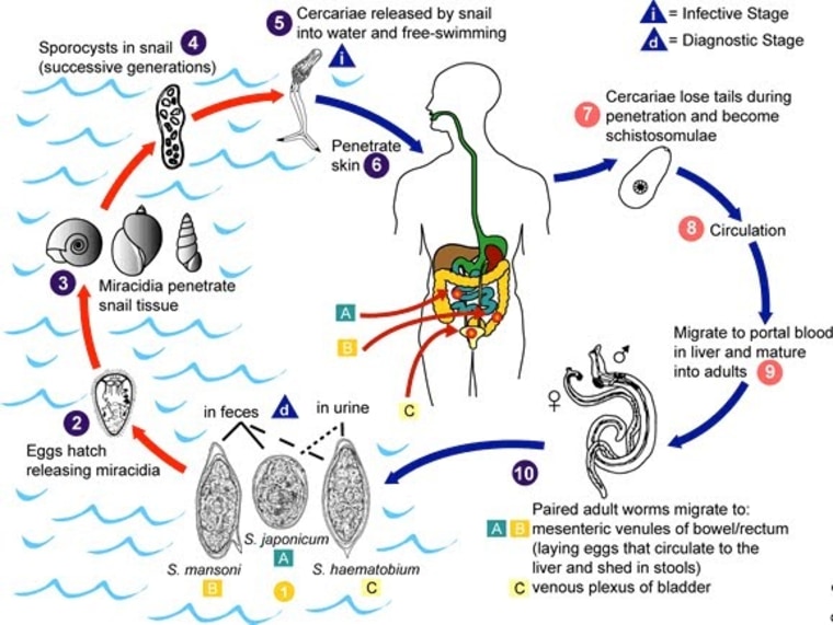
The researchers said they could only speculate on what may have caused of the young man's cancer, but it may have been as a result of environmental carcinogens such as smoke from wood fires, or due to genetic factors, or from an infectious disease such as schistosomiasis, which is caused by parasites.
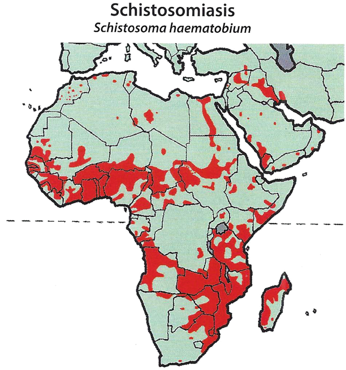
Schistosomiasis would be a plausible explanation, they said, since the disease has plagued inhabitants of Egypt and Nubia since at least 1500 BC and is now recognised as a cause of bladder cancer and breast cancer in men.
The species of schistosomes common to Africa is S. haematobium. Of course, like many parasites schistosomes cause a variety of health problems to their human hosts who are actually not the primary host but the secondary or tertiary hosts. In the case of S. haematobium, the adult worms congregate in the bladder which is used as their breeding ground where they mate and lay eggs that pass out the urine. The problem is that this congregation of worms causes inflammation and other bladder issues.
Another issue that schistosomes and other parasites cause are other diseases due to immune suppression. In order to evade immune response from the host, the parasite would release antigens that suppress the immune system which leads to other issues in the long run such as cancers like carcinoma which was the type found in the Nubian mummies discussed above. It's probable if not likely their cancers were caused by schistosomes.
Posted by Djehuti (Member # 6698) on :
Anemia seemed to have been prevalent among ancient Egyptian children.
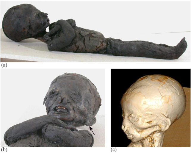
Anemia found to be common in ancient mummified Egyptian children A team of paleopathologists and medical experts from Germany, the U.S. and Italy has found that anemia was common in ancient Egyptian children who had been mummified. In their study, reported in the International Journal of Osteoarcheology, the group subjected multiple mummified remains of children from ancient Egypt to computed tomography scans to study their skeletons.
The research team focused their efforts on children from that time that had died before reaching adulthood and who had then been mummified. Mummifying the children allowed their remains to be preserved in ways not possible with those who were simply buried. But modern study does not allow unwrapping the dressings used in the mummification process; thus, researchers have to use modern machines to peer through the dressings to learn more about what is inside.
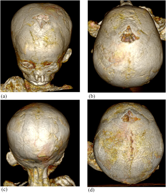
In this new effort, the researchers ran full-body CT scans on 21 child mummies (between the ages of 1 and 14) in order to study the entire skeleton. In so doing, they found evidence of pathological enlargement of the cranial vaultthe part of the skull that holds the brainin seven of the children. Such enlargement is typically associated with anemia.
..Anemia is generally caused by malnutrition. It leads to reduction in production of red blood cells, which means that not enough oxygen can be carried to the brain and other parts of the body including the bones. Those with anemia also typically suffer from other problems, as well, such as iron deficiency, bleeding in the gastrointestinal tract, inflammation and chronic infections due to a weakened immune system. It was not possible to tell from the CT scans if anemia contributed to the deaths of the children, but the research team suggests it was, at the very least, a contributing factor.
The team also found a child who had suffered from thalassemia, in which the body cannot produce hemoglobin, and who also had an enlarged tongue. That child lived less than a year, almost certainly succumbing to the many symptoms associated with the disorder.
While a common cause of anemia in impoverished regions and especially in ancient times was malnutrition or parasitic infections, the dysfunction could also be caused by blood cell disorders like Thalassemia or sickle cell.
Researchers used computed tomography (CT) scans to peer non-invasively through the mummies' dressings and discovered that one-third of them had signs of anemia; they found evidence of thalassemia in one case, too.
"Our study appears to be the first to illustrate radiological findings not only of the cranial vault but also of the facial bones and postcranial skeleton that indicate thalassemia in an ancient Egyptian child mummy," the team writes in their published paper.
Paleopathologist Stephanie Panzer and her colleagues from Germany, the US, and Italy, suggest that anemia was likely common in ancient Egypt, and it was probably caused by factors such as malnutrition, parasitic infections, and genetic disorders, which still cause the health problem today.
Researchers have even speculated that Tutankhamun died of sickle cell disease, a cause of anemia. However, as the researchers of this new study explain, "the direct evidence of anemia in human remains from ancient Egypt is rare."
Anemia is a condition where the body lacks enough healthy red blood cells to carry oxygen to the body's tissues. As Panzer and colleagues studied child mummies, the remains are more likely to show signs of anemia than adult mummies, due to their early death.
Whether or not anemia played a role in each of the children's deaths could not be determined from the CT scans, but the research team believes it is likely to have contributed. They also looked for signs of diseases that could have caused the anemia.
When ancient humans were mummified, their bodies were preserved in ways that kept more information than those buried. Although modern science doesn't let researchers remove the wrappings used in the mummification process, they often use scans to 'look' through the wrappings and see what's inside.
It's funny that they mention the 2010 finding by a German team that King Tut may have died from sickle cell disease.
There was a genetic study done in 1999 on a sample of 6 late predynastic mummies and half of them tested positive for HbS and the disease is still endemic to Egypt today especially among inhabitants of the western oases: Marin, A., Cerutti, N. and Massa, E.R. (1999) Use of the amplification refractory mutation system (ARMS) in the study of HbS in predynastic Egyptian remains. Boll. Soc. Ital. Biol. S
King Tut died of blood disorder: German researchers But the German researchers said in a letter published online Wednesday by the Journal of the American Medical Association that closer scrutiny of his foot bones pointed to sickle cell disease, in which red blood cells become dangerously misshaped.
"We question the reliability of the genetic data presented in this (the Egyptian) study and therefore the validity of the authors' conclusions," the letter said.
"(The) radiological signs are compatible with osteopathologic lesions seen in sickle cell disease (SCD), a hematological disorder that occurs at gene carrier rates of nine percent to 22 percent in inhabitants of Egyptian oases."
From what I remember the Egyptian team has yet to conduct tests to confirm the presence of HbS.
Posted by Djehuti (Member # 6698) on :
For the sake of saving bandwidth space I'll post this here:
Egyptian and Greco-Roman family graves expected to provide new insight into ancient diseases and burials Dozens of family tombs containing mummies and artefacts have been discovered near the city of Aswan in southern Egypt.
An Egyptian-Italian archaeological mission unearthed 33 tombs while working near the Aga Khan Mausoleum west of Aswan, the Egyptian Ministry of Tourism and Antiquities announced on Sunday.
The tombs date from the Late Period of Ancient Egypt (664-332BC), the Ptolemaic period when the country was ruled by a Greek-speaking dynasty (305-30BC), and the Roman period (30BC-641AD).
The tombs, some of which still contain mummified remains and funerary objects, are expected to provide new insight into the history of the Aga Khan area and diseases that afflicted its ancient inhabitants.
"This discovery adds new history to the Aga Khan area," said Dr Mohamed Ismail Khaled, Secretary General of the Supreme Council of Antiquities.
Dr Ayman Ashmawy, Head of the Egyptian Antiquities Sector at the Supreme Council of Antiquities, said preliminary studies on the mummified remains suggest 30 per cent to 40 per cent of those buried in the tombs died young, ranging from newborns to adolescents.
The tombs vary in architectural design, with some featuring vaulted entrances preceded by open courtyards surrounded by mud-brick walls while others are carved directly into the mountain rock.
Among the discoveries within the tombs are several mummies, including those of an adult, possibly a woman, and a child who may have died between the ages of one and two. The two bodies were found still adjoined inside a stone sarcophagus, a mystery the mission plans to investigate further.
Other finds include remnants of coloured cartonnage, clay and stone figurines, wooden coffins and offering tables.
Dr Abdel Moneim Saeed, General Supervisor of Aswan and Nubia Antiquities and director of the Egyptian side of the mission, suggested the middle class of Aswan Island's inhabitants were buried in this part of the necropolis, while the upper part was designated for the upper class.
Advanced technology, including X-ray analysis, has been used to study the discovered mummies, revealing details about their facial features, sex, age at death and the presence of organic diseases.
Dr Patrizia Piacentini, professor of Egyptology at the University of Milan and director of the Italian side of the mission, noted that preliminary studies on the mummies indicated some suffered from infectious diseases, bone disorders, anaemia, malnutrition, chest disease, tuberculosis and osteoporosis.
Some were found to have died at an advanced age with severe bone disease.
The mission is expected to continue its work at the site, aiming to discover more about the ancient inhabitants of the area.
Advanced technology, including X-ray analysis, has been used to study the discovered mummies, revealing details about their facial features, sex, age at death and the presence of organic diseases.
Dr Patrizia Piacentini, professor of Egyptology at the University of Milan and director of the Italian side of the mission, noted that preliminary studies on the mummies indicated some suffered from infectious diseases, bone disorders, anaemia, malnutrition, chest disease, tuberculosis and osteoporosis.
Some were found to have died at an advanced age with severe bone disease.
The mission is expected to continue its work at the site, aiming to discover more about the ancient inhabitants of the area. Posted by Archeopteryx (Member # 23193) on :
I see that a new book has been published about ancient Egyptian medicine. I have not read it yet but it looks interesting.
In the presentation they mention that medical treatment often was accessible beyond the affluent class, something that not all countries in todays world can offer.
quote:Medicine and Healing Practices in Ancient Egypt: A Comprehensive Exploration
Ancient Egypts healthcare system, described as advanced and successful by researchers Rosalie David and Roger Forshaw in their new book, Medicine and Healing Practices in Ancient Egypt, offers a unique perspective on the practices that thrived in the land of the pharaohs over three millennia.
Affiliated with the University of Manchester in the United Kingdom, the authors present a people-focused interpretation, shedding light on the interaction between healthcare providers and their patients.
The comprehensive study delves into the intricacies of ancient Egyptian medicine, highlighting pharmaceutical treatments involving minerals, plant ingredients, and animal parts. Basic surgeries, pragmatic remedies like bandaging broken limbs, and, to a lesser extent, magical treatments were universally available across all societal levels.
Professor Rosalie David, an emeritus professor of biomedical Egyptology, says: The Egyptian healthcare system was advanced and successful, not least for devising innovative ways to treat snake bites and save lives. The researchers point out that despite the absence of a complete picture of ancient Egyptian life, evidence suggests that individuals had some autonomy in choosing their healthcare providers.
Inscriptions reveal the dual religious and secular roles of physicians, who not only provided medical treatment but also served as priests in healing-associated deities temples. Payment for care was likely based on affordability, making treatments accessible beyond the affluent class. Physicians and health workers, including midwives, visited patients homes, while temple precincts provided centralized locations for treatments and therapy.
The ancient Egyptians did not categorize diseases; instead, their medical documents focused on individual case studies, listing symptoms and potential outcomes. Infectious diseases were sometimes attributed to deities or perceived enemies, requiring prayers, rituals, or magical treatments. Mental illnesses were recorded based on symptoms rather than specific diseases.
Notably, the ancient Egyptians displayed an enlightened attitude toward deformities and disabilities, unlike their Greek counterparts. Individuals with disabilities were not excluded from working in temples, and specific careers were designated to provide livelihoods for diverse groups. This progressive approach extended to old age, where cosmetic treatments were used to combat the appearance of aging.
Addressing the prevalence of snake and scorpion bites, the researchers highlight the Brooklyn Papyrus, dating back to around 450 BCE, as one of the oldest writings about medicine. The document outlines various snakes, their habitats, symptoms of bites, and deity associations, offering treatments ranging from practical interventions to magical incantations. The use of natron, a naturally occurring compound, showcased the Egyptians practical knowledge in treating such ailments.
Despite an average life expectancy similar to other ancient societies, approximately 90 percent of the adult population in ancient Egypt did not live beyond the age of fifty. Notable exceptions, like Pharaoh Ramesses II, underscored the rarity of individuals reaching old age.
The enduring legacy of ancient Egyptian healthcare is evident in various aspects of modern Western medicine. Techniques for treating dislocated jaws, recorded in the Edwin Smith Papyrus around 1600 BCE, remain in use. The Egyptians pioneering use of sutures and medical instruments like scalpels is also acknowledged.
While surgical treatments were limited, evidence from medical papyri, such as the Ebers Papyrus, detailed surgical procedures like incisions for swellings. Bandages and wound coverings made of linen were common, showcasing early wound management practices. The Ebers Papyrus also discusses medicinal formulations for treating burns.
The researchers underscore that despite appearing unsophisticated by twenty-first-century standards, ancient Egyptian medicine demonstrated evidence-based practices four millennia ago. The centrality of the patients role and the enlightened attitude toward deformities and disabilities constitute the Egyptians enduring legacy to the modern world.
More information: David, R., & Forshaw, R. (2023). Medicine and Healing Practices in Ancient Egypt. doi:10.3828/9781837644292
^ One reason for the Egyptians' advanced medicine was their extensive knowledge of anatomy which was likely based on experiments done on animals, particularly livestock and perhaps even traditions of aruspicina (divination using animal entrails) and other organoscopy (divination using organs in general). This is a practice found in other parts of Africa. No doubt there was practice of surgery on live patients based on inference from that of animals. Mind you the cutting open of a dead human (or other sacred animal) was considered offensive which is why ritually during mummification there was one type of priest would perform the incision on the body after which he had to be ritually chased away and insulted, and in some cases had small stones thrown at him. There are reports from the Romans and later Europeans in Medieval times of Moors and other West African having surgical knowledge as well.
African Tribes that Pioneered Surgery Lets take a journey through time to uncover the surgical pioneers among African tribes who, long before modern medicine, crafted their own surgical traditions. These practices showed a profound understanding of the human body without a textbook. This was knowledge passed down to generations. From C-sections to amputations, African tribes were performing medical miracles in the context of today.
Another interesting fact is although the Egyptians and other Africans did not know about germ theory they believed disease causing spirits had affinity to dirt and impurity, which is why whenever a cut was made on a living person especially surgeries, they made use of antiseptics to clean the area and made sure the surgery was done in a closed setting that was as clean as possible often waving the air around using a flaming torch or bundle of burning leaves to cure the air around.
Posted by Djehuti (Member # 6698) on :
The tumor, known as a teratoma, was discovered alongside a ring that may have been thought to have magical powers to defend against pain.
The discovery in the North Desert Cemetery of Amarna, Egypt, could shed light on how ancient Egyptians dealt with disease, Gretchen R. Dabbs, a professor of bioarchaeology at Southern Illinois University Carbondale, told Insider.
The remains belonged to a woman who died at the age of between 18 and 21, according to the case study, published in the International Journal of Paleopathology.
We don't know what killed the woman, according to Dabbs, who was the lead author of the study. However, she may have been suffering from pain from the teratoma in her ovaries.
Teratomas are a very rare type of tumor that still occur today. They are made up of a type of cell that can sometimes spontaneously turn into other parts of the body, which means they can sprout hair, teeth, bones, or muscles.
The tumors are mostly benign but can sometimes cause pain and issues with conception. Even today, they often go completely unnoticed.
It is possible this teratoma was bothering the ancient Egyptian woman 3,000 years ago. She was buried with rings on her left hand, which were placed above the tumor. While the placement of the hand is customary for such burials, one of the rings may not be: this one carried the symbols of Bes, a deity commonly associated with childbirth, fertility, and protection.
If the rings were placed above the tumor, would they have had any actual effect on it?
Posted by Djehuti (Member # 6698) on :
^ The rings were suppose to be amulets with magical/blessing affects to ease pain. Whether it worked as a form of psycho-placebo or not we'll never know.
I suspect the gynocologists of her time who were most likely priestesses did not want to perform surgery to remove it since such a procedure on the uterine wall was very risky so all they could offer was palliative care.